Spine Anatomy
The human spine is a complex and vital structure that provides structural support, protects the spinal cord, and enables movement. It is divided into five distinct regions, each with specific functions.
Key Anatomical Components
- Vertebrae: The 33 individual bones that stack to form the spinal column and protect the spinal cord.
- Intervertebral Discs: Acts as shock absorbers between the vertebrae, providing flexibility.
- Facet Joints: Joints between the vertebrae that allow for twisting, turning, and bending.
- Spinal Canal: The central tunnel formed by stacked vertebrae that houses and protects the spinal cord.
- Nerve Roots: Nerves that branch off the spinal cord to reach different parts of the body.
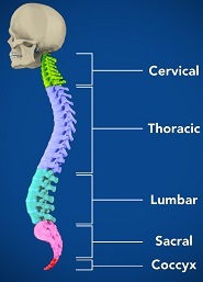
Regions of the Spine
- Cervical Spine (Neck): The top 7 vertebrae (C1–C7) that support the skull.
- Thoracic Spine (Upper Back): The 12 vertebrae (T1–T12) that connect to the rib cage.
- Lumbar Spine (Lower Back): The 5 largest vertebrae (L1–L5) that bear the body's weight.
- Sacrum: Triangular bone at the base connecting the spine to the pelvis.
- Coccyx (Tailbone): The very bottom of the spinal column.
Understanding the normal anatomy of the spine is essential for identifying conditions and injuries that may affect your mobility and comfort.